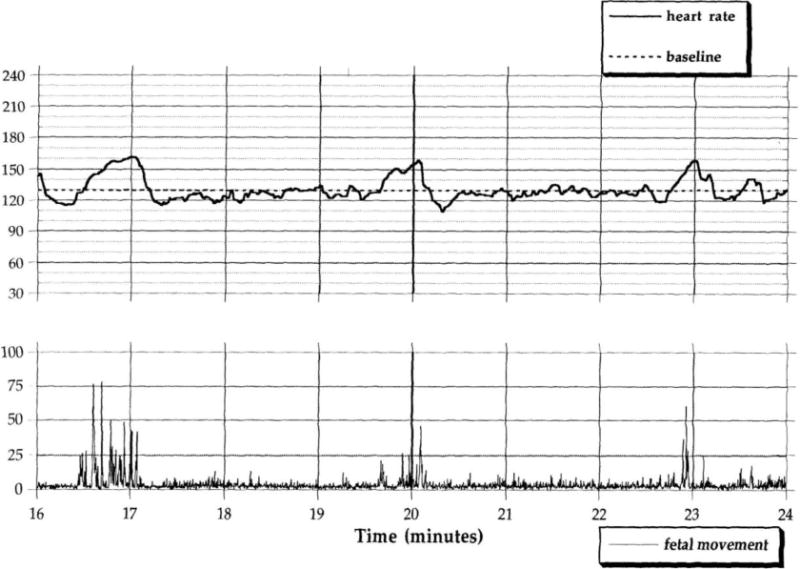
Figure 1.

Examples of 8 minute segments of digitized fetal heart rate and fetal movement data in a 36 week fetus. The upper figure reflects a period of fairly brief movements (i.e., at minutes 16.5, 20, and 23) against a background of quiescence associated with self-limited accelerations in fetal heart rate. The lower figure shows periods of more persistent, unregulated movement and highly variable fetal heart rate.
