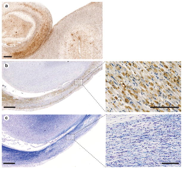
Figure 3.
Neuroinflammation and white matter degeneration after TBI. a | Extensive reactive microglia (immunostained with antibody CR3/43) in the hippocampus and adjacent sulci of a 65-year-old male with dementia pugilsitica. Scale bar 1 mm. b | Extensive CR3/43-reactive cells with an amoeboid morphology, indicative of macrophages, observed in the atrophic corpus callosum of a 37-year-old male 4 years following a single severe TBI. Scale bar 1 mm for main image (left) and 100 μm for high-magnification image (right). c | Adjacent section to b, stained with Luxol fast blue, indicating chronic white matter change and loss of myelin. Scale bar 1 mm for main image (left) and 100 μm for high-magnification image (right). Abbreviation: TBI, traumatic brain injury.

