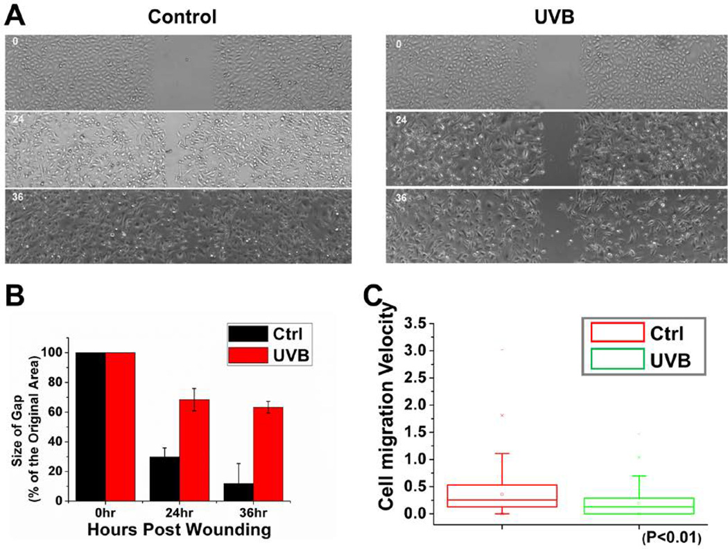
Figure 2. Cell migration defects in UVB-treated keratinocytes.
(A) Migration of confluent monolayers of mouse keratinocytes cultured from control and UVB treatment was assessed by in vitro scratch-wound assays. (B) The kinetics of in vitro wound healing was quantified. Wounding areas were measured using ImageJ software and expressed as the percentage of the original area size with the bar graph (P < 0.01). A representative experiment is shown. (C) Movements of individual keratinocytes were traced by videomicroscopy. Migration tracks of multiple cells for each group (control or UVB treated) are shown here as scatter plots.

