
Figure 2.
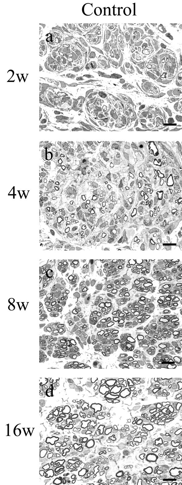
Regeneration in control xenografts at 2 (a), 4 (b), 8 (c), and 16 (d) weeks after surgical procedures. One-micrometer-thick, toluidine blue-stained cross-sections taken ∼1 mm proximal to the distal graft host junction are shown. Sural nerve was obtained from the normal donor, and nerve xenografts were generated as described in Materials and Methods. By 2 weeks, many myelinated axons, predominantly in the 2-4 μm size range, had regenerated into the graft. The density of all fiber sizes increased at 4 and 8 weeks, but a relative increase in the number of very small (<2 μm) MFs was noted. At 16 weeks, there was a substantial increase in the number of large (>4 μm)-diameter fibers (for histograms, see Fig.4a-d). Scale bars, 10 μm.