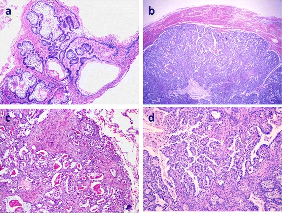
Fig. 2.

Histological findings of MA. (a): The locally recurrent tumor showed a solid/multicystic-type ameloblastoma. The tumor nests consisted of stellate cells with peripheral palisading. The stroma was fibrous. HE × 40. (Case 2). (b): The pulmonary metastatic lesion was more cellular, with a clear margin between the lesion and the surrounding lung tissue. HE × 40. (c): Spindle cells and oval cells comprised the nests and intercrossed the margin with glandular structures. (d): Many glandular/papillary structures were observed, among which were sheets of spindle cells with local squamous metaplasia. Cytological atypia was absent. HE × 100
