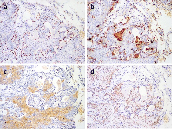
Fig. 3.

Immunohistochemical staining in MA. (a) and (b): The lining epithelial cells of the gadular structures are TTF-1+ and PE10+, indicating the residual alveolar epithelial cells (×100). (c) Stellate cells are CK10/13+ (×100). (d) The spindle cells are also p63+ (×100)
