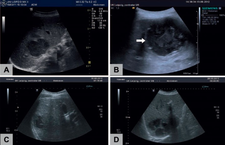
Fig. 2.
Ultrasound morphology of LA (B mode): PLA frequently show an inhomogeneous hypoechoic pattern (A) with a thickened, edematous wall (B, arrow). Blurred, irregular borders (C) and septa (D) can also be observed. Morphology cannot discriminate between pyogenic (A, B, D) and amebic (C) abscesses.

