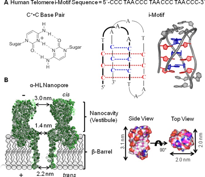
Figure 1.

Structures of the human telomere i-motif and the α-hemolysin nanopore. (A) The C+•C base pair scheme, the human telomere i-motif line structure, and cartoon drawing based on pdb 1ELN.9 (B) Space filling model of the α-hemolysin nanopore (pdb 7AHL)10 and the human telomere i-motif along with their dimensions for comparison.
