
Figure 5.
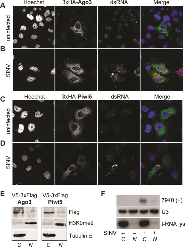
vpiRNA biogenesis occurs in the cytoplasm. Localization of 3xHA-tagged Ago3 (A,B) and Piwi5 (C,D) in uninfected (A,C) and SINV (parental virus) infected Aag2 cells (B,D) as determined by confocal microscopy. Hoechst staining indicates the nuclei. Infected cells were identified by a strong cytoplasmic dsRNA staining, which can be clearly distinguished from low-level background staining in non-infected cells. (E) Western blot analysis of V5–3xFlag-tagged Piwi5 and Ago3 in the cytoplasmic (C) and nuclear (N) fraction of Aag2 cell lysates. Tubulin and Histone 3 dimethylated at lysine 9 (H3K9me2) were used as cytoplasmic or nuclear markers, respectively. (F) Northern blot of vpiRNA 7940 (+) in nuclear and cytoplasmic fractions of Aag2 cells. U3 RNA and t-RNA lysine serve as nuclear and cytoplasmic marker, respectively.