Fig. 2.
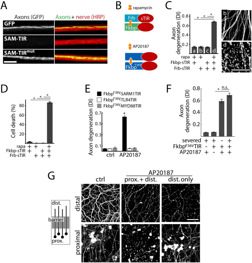
Axon degeneration and neuronal death induced by sTIR dimerization. A) Micrograph showing motor nerves of third instar Drosophila larvae. M12-Gal4 drives expression from mCD8-GFP (green) alone or with either UAS-SAM-TIR or UAS-SAM-TIRmut in single motor axons in each nerve (red=HRP). UAS-SAM-TIR expression caused axon loss in 49/49 nerves as shown; whereas SAM-TIR with a disruptive TIR mutation led to degeneration in 0/70 nerves (χ2=119; p<0.001); scale bar=10 micrometers. B) Schematic showing sTIR dimerization by rapamycin or AP20187. C) Effect of sTIR, dimerized sTIR, and rapamycin on axon degeneration. α-Tubulin stained axons correspond to bars b and d. D) Effect of sTIR dimerization on neuronal viability quantified by ethidium homodimer exclusion after 24 hours. E) Effects of dimerization of sTIR or TIR domains of MYD88 or TLR4 on axon degeneration. F) Effects of sTIR dimerization on degeneration of Sarm1−/− axons physically disconnected from cell bodies. G) Left: diagram of axons growing through a diffusion barrier into an isolated fluid compartment. Right: micrographs of isolated distal axon segments after application of AP20187 globally or selectively to distal axons. Scale bar = 50 micrometers. Error bars = SEM; * p < 0.01; one-way ANOVA with Tukey’s post-hoc test.

