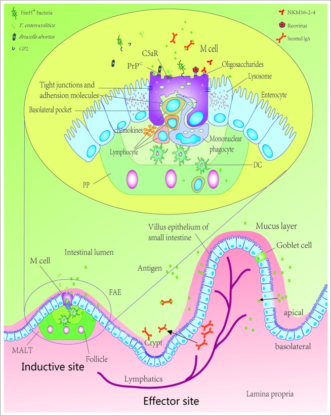
Figure 1.
Schematic diagram of intestinal epithelium showing M cells, PPs, intestinal epithelial cells and translocation mechanisms of several kinds of antigen. Gut-associated mucosal immune system has 2 parts, including inductive sites and effector sites. The inductive sites are mainly composed of MALT, and the effector sites are primarily constituted by the lamina propria of various mucosae, stroma of exocrine glands, and surface epithelia. In the FAE, luminal antigens are transported across the epithelium by M cells, where primary immune responses can be induced, and then phagocytosed by DCs present in the dome of the PPs, delivered to the mesenteric lymph nodes, which can directly prime T-cell responses to antigens. DCs also sample antigens through enterocytes. sIgA, secreted by mature plasma cells in the lamina propria, from epithelial cells into the gut lumen, may have a controlling role in bacterial persistence and uptake. M cells can transport bacteria, viruses, parasites and non-infectious particles through the apical membrane to the basolateral surface, exploiting many molecules and proteins to recognize and transport antigen. M cells lack microvilli and possess basolateral pockets containing mononuclear phagocytes and lymphocytes, which contribute antigens transported to the underlying tissues. PP, Peyer's patches; DC, dendritic cell; GP2, Glycoprotein 2; PrPC, cellular prion protein; C5aR, C5a receptor; FAE, follicle-associated epithelium; MALT, mucosa-associated lymphoid tissue.

