Figure 1.

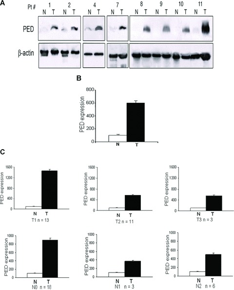
PED expression is increased in human lung cancer. (A) Western blots showing expression of PED in tumour (T) and adjacent normal (N) lung tissue from some of the 27 NSCLC-affected patients (Pt). β-Actin was used for the loading control. (B) Graph of densitometric analysis. Mean ± SD PED expression of all the tumour samples (T) normalized to β-actin and expressed as percentage respect to the adjacent normal tissue (N). PED was >6-fold higher in cancer tissue compared to normal areas. (C) PED expression during T1, T2, T3 and N0, N1 and N2 stages of the disease. PED expression is greater during the initial stages (T1 and N0) of the disease compared with the T2 and N1 lesions.
