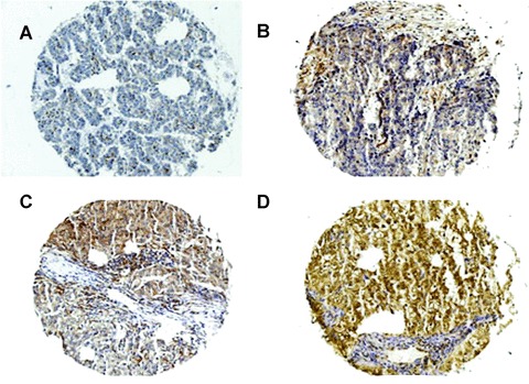
Figure 2.

PED expression levels in NSCLC cancer samples. Immunohistochemical analysis of paraffin-embedded NSCLC sections labelled with anti-PED antibody (1:5000) and revealed by secondary, biotinylated antibody. (A) Negative; (B) mild; (C) moderate and (D) strong staining.
