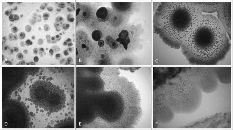
Figure 2.
Development of L-form growths in semisolid agar during the second phase/week of cultivation. Light microscopy of blood isolates (No1; 34; 37, 48; 91; 96): typical “fried eggs” colonies of different size, consistence and density (A, B, C, D); formation of biofilm with gliding motility at the periphery (E, F). Magnification: 400x

