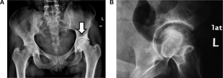
Figure 1.

(A) Plain radiography (anteroposterior (AP) view of both hip joints) and (B) pelvic radiography (close oblique view of the left hip).
Notes: Plain radiography of the hip joints: AP view and oblique view of the left hip showing heterogeneous matrix of the left femoral head giving geographical appearance with mixed sclerosis and lucent areas as well as relative mild narrowing of the ipsilateral joint space representing AVN-related changes.
