Figure 2.
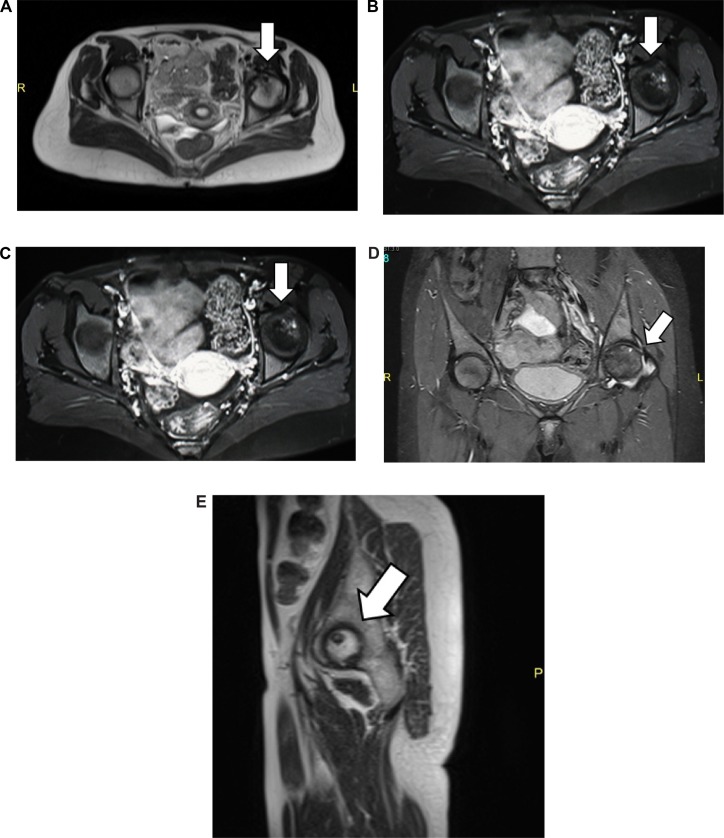
MRI of both hip joints: (A) axial view of the T2WI sequence, (B) axial view of the T1WI sequence post-contrast (gadolinium DTPA) intravenous administration, (C) coronal view of the T2WI sequence, (D) coronal T2 STIR sequence, and (E) sagittal view of the left hip joint T2WI sequence.
Notes: MRI examination of the hip joints before and after intravenous administration of gadolinium. Axial, coronal, and sagittal views of the hip joints T1, T2, and STIR showing multiple subarticular areas of abnormal signal intensity within the head left femur, mainly at the superoanterior medial aspect showing low T1 signal intensity and non-homogenous mixed T2-fat suppression with mild enhancement. Associated mild degenerative changes and thickened synovium are noted, consistent with stage III–IV AVN. No signs of AVN on the right hip were detected.

