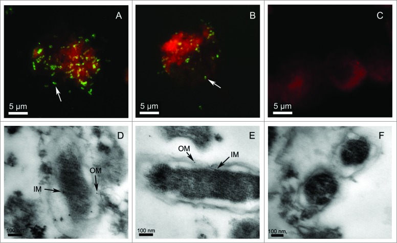
Figure 2.
Microscopy analysis of YbgF and TolC in R. rickettsii cells. Rickettsia rickettsii were cultured in Vero cells and incubated with antibodies against rYbgF (A) or rTolC (B), or with naïve (C) serum. Secondary antibodies conjugated to fluorescein isothiocyanate (FITC) were then applied. The white arrows indicate YbgF or TolC in R. rickettsii cells. TEM analysis of R. rickettsii in Vero cells involved staining with antibodies against rYbgF (D), rTolC (E), or with naïve (F) serum. A goat anti-mouse IgG labeled with colloidal gold particles was then added to samples. The black arrows indicate the locations of YbgF or TolC in the inner membrane (IM) and outer membrane (OM) of R. rickettsii cells.

