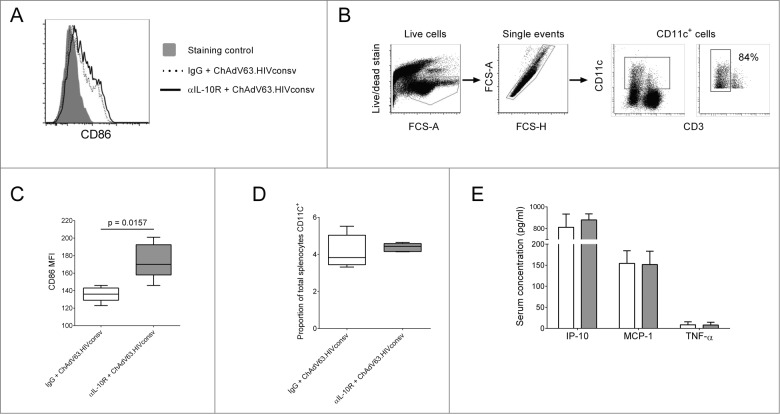
Figure 3.
The effect of IL-10R blockade on innate responses to ChAdV63.HIVconsv. (A) Representative histogram showing CD86 expression on CD11c+ cells 24 hours post-immunization in mice immunized with ChAdV63.HIVconsv in combination with either anti-IL-10R antibody (solid line) or an irrelevant isotype control antibody (dashed line). (B) Gating strategy for the identification of CD11c+ cells and representative plot showing the percentage of CD11c+ cells that are CD3- (far right plot). (C) CD86 expression on CD11c+ cells 24 hours post-immunization in mice immunized with ChAdV63.HIVconsv in combination with either anti-IL-10R antibody (gray bar; n = 5) or isotype control antibody (white bar; n = 5). Statistical significance was determined using the Mann Whitney test. (D) The proportion of splenocytes expressing CD11c 24 hours post-immunization in mice immunized with ChAdV63.HIVconsv after receiving anti-IL-10R (gray box; n = 5) or isotype control antibody (white box; n = 5). (E) Concentration of IP-10, MCP-1 and TNF-α in serum collected 24 hours post-immunization from mice immunized with ChAdV63.HIVconsv in combination with anti-IL-10R (gray bars; n = 10) or isotype control antibody (white bars; n = 10). Data are representative of 2 independent experiments.

