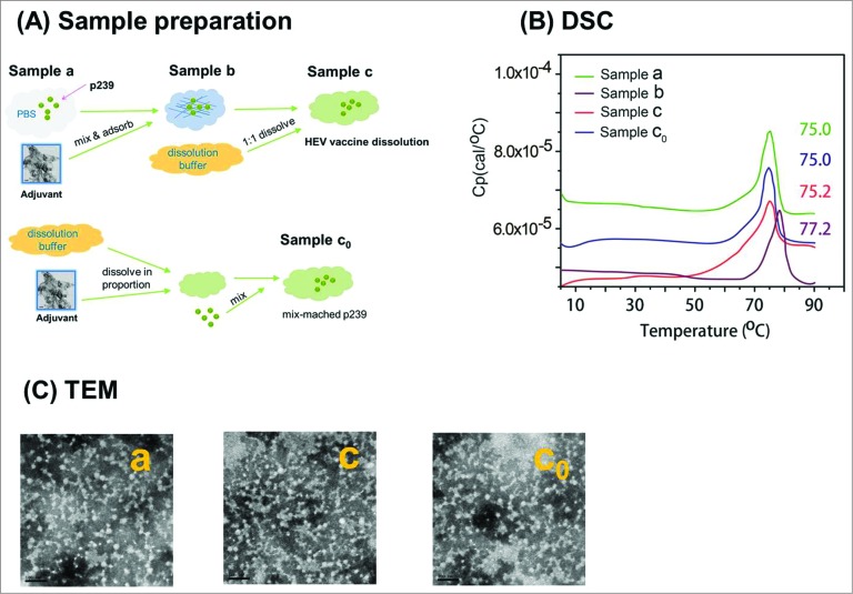
Figure 1.
The sample preparation and consistent profiles of p239 vaccine antigen particles detected by DSC and TEM. (A) The samples were prepared according to the flow diagram. The process shows the preparation of p239 VLPs in Samples a, c, and c0 that were used for multiple analyses, with Sample c0 being the matrix-matched control for Sample c. (B) Comparable thermal stability as shown by DSC profiles – similar transition temperatures (between 75°C to 75.2°C) were obtained for p239 VLPs in Samples a, c and c0, and a slightly higher transition temperature (77.2°C) was observed in absorbed p239 VLPs in Sample b. (C) Morphology of p239 VLPs were examined using transmission electron microscopy (TEM). The spherical particles of native (Sample a) and aluminum-dissolved p239 VLPs (Samples c and c0) presented similar morphology (Bar = 100 nm).

