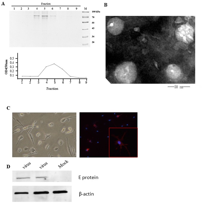
Fig 2. Purification of DENV-2 particles from insect cells and infection of MDDC.
(A) DENV-2 virus was concentrated and purified via sucrose density gradient centrifugation. Fractions from the gradients were analyzed by SDS-PAGE, and dengue virus was mainly found in fractions4, 5 and 6 after centrifugation (upper panel). Double anti-DENV-2 antibody ELISA results were consistent with electrophoresis(lower panel).(B) Purified mature DENV-2 virions were negative stained and observed via transmission electron microscopy. Mature dengue virions were approximately 50 nm in diameter, surrounded by lipid bilayers, and had‘‘smooth” regions on their outer membranes. (C) Monocytes isolated from PBMCs were treated with 25 ng/ml IL-4 and 50 ng/ml GM-CSF for 7 days and infected with DENV-2 at an MOI = 0.1 for 48 hours. Two days after infection, the cells were permeabilized and analyzed for DENV-2 E protein expression using a 4G2 antibody. Nuclei were stained with DAPI (blue). (D) The expression of DENV-2 E was demonstrated by western blotting using anti-DENV-2 hyperimmune serum.

