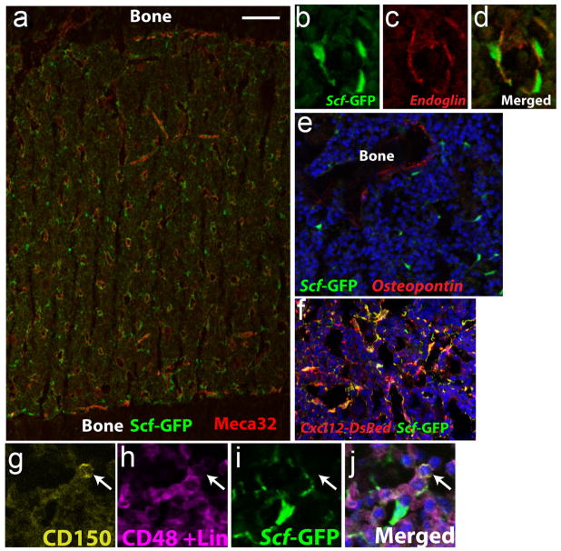
Figure 2. HSCs and their niche cells surround sinusoids throughout the bone marrow.
a. Sections through the bone marrow of Scfgfp/+ mice show that HSC niche cells (green) include mesenchymal stromal cells and endothelial cells that surround sinusoids and potentially other blood vessels throughout the bone marrow64. b–d. High magnification shows that Scf-GFP overlaps with the endothelial marker endoglin but also extends beyond the endoglin on the abluminal side of the sinusoids, indicating expression by mesenchymal stromal cells. e. Scf-GFP is not expressed by osteopontin+ bone lining cells around trabecular bone, but is expressed by some nearby perivascular cells. f. Cxcl12-DsRed exhibits a similar expression pattern, primarily by perivascular mesenchymal cells and endothelial cells around sinusoids throughout the bone marrow, in a pattern that strongly overlaps with Scf-GFP in Cxcl12DsRed/+; Scfgfp/+ mice17. g–j, Cells that are CD150+ (g) and CD48 and Lineage marker negative (h) are usually found immediately adjacent to Scf-GFP+ perivascular cells (i) in the bone marrow (see j for merge). Images are from references17,64.

