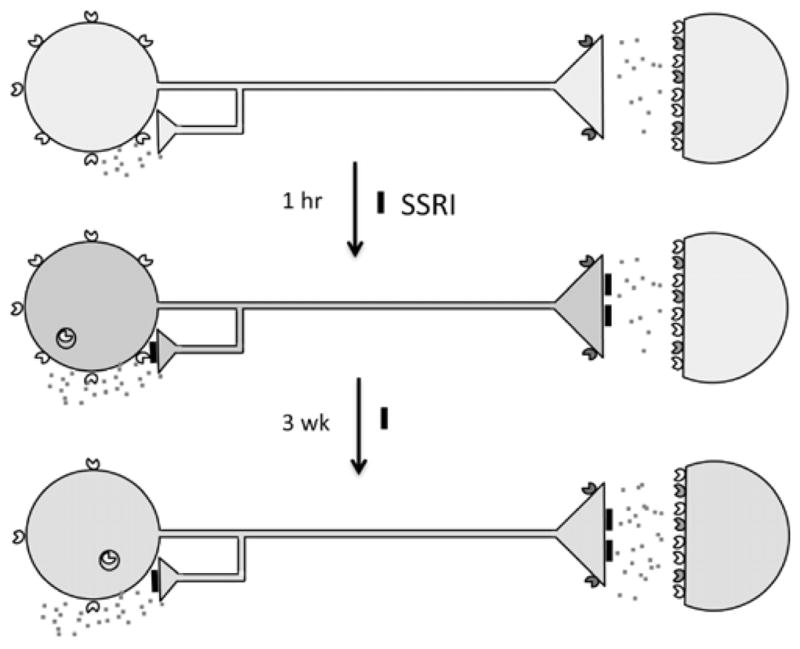
Fig. 1. Adaptive changes in 5-HT neurons upon antidepressant treatment.

In major depression, 5-HT neurons are thought to have reduced neurotransmission (pale blue). 5-HT1A autoreceptors (yellow carets) are expressed presynaptically on 5-HT neuron cell bodies and dendrites (at left); 5-HT1B receptors (green carets) are presynaptic at the nerve terminal; 5-HT1A heteroreceptors and other 5-HT receptors are expressed postsynaptically (colored carets). The 5-HT1A receptor couples to Gi/Go protein to inhibit adenylyl cyclase (AC), open potassium channels (K+) to hyperpolarize membrane potential and reduce firing rate. Acutely, SSRI antidepressants inhibit (black blocks) the 5-HT transporter increasing synaptic 5-HT (red specks), leading to activation of 5-HT1A autoreceptors which inhibit 5-HT neuronal firing (blue), and transiently internalize. After 3 weeks of SSRI treatment, 5-HT1A autoreceptors desensitize and receptor levels are reduced, dis-inhibiting 5-HT neuronal firing and enhancing 5-HT neurotransmission (green).
