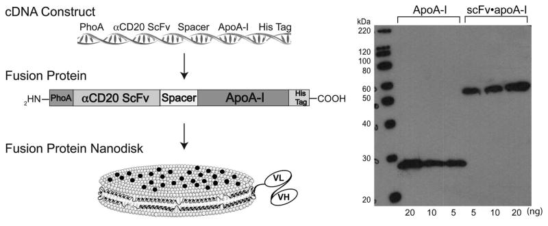
Fig. 1.

αCD20 scFv•apoA-I design, construction, expression and characterization. (Left) Schematic depicting αCD20 scFv•apoA-I chimera cDNA and protein. Also depicted is the fusion protein as the scaffold component of a ND (the black circles represent curcumin embedded in the phospholipid bilayer). (Right) SDS-PAGE immunoblot analysis of αCD20 scFv•apoA-I fusion protein. Samples were electrophoresed on a 4%–20% acrylamide gradient SDS slab gel under reducing conditions, transferred to PVDF membrane, and probed with anti-apoA-I. Left lane, molecular weight standards; center lanes, recombinant human apoA-I; right lanes, recombinant αCD20scFv•apoA-I fusion protein. Protein load (ng per well) are indicated.
