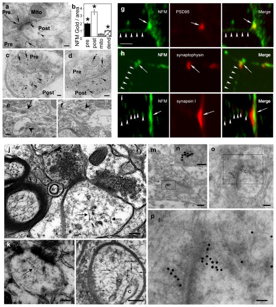
Figure 1.
Presence of NFM proteins in synapses and 10nm NF polymers in synapses. Post-embedding EM using anti-NFM antibody (RMO-44)(a) shows immunogold decoration of pre-synaptic (a,” Pre”, arrows) and post-synaptic regions (a, “Post”, arrowheads). Mitochondria (Mito), as a background control, exhibit little or no labeling. (b) Morphometry of NFM-gold particles shows 8-fold more immunogold in post-synaptic regions (P<0.0001) per unit area than in mitochondria and 4-fold more than in pre-terminal dendrites where the presence of NFM is established (P<0.0001). Gold particles in the pre-synaptic region are 4-fold more numerous than in mitochondria. (c, d) Two examples illustrating the greater immunogold labeling in post-synaptic regions than in pre-synaptic regions (” Pre”, arrows and “Post”, arrowheads). Negative controls without primary antibody (e) or with primary antibody pre-absorbed (f) show negligible labeling. Arrow points to a dendrite in (e) and arrowheads to synapses in (e, f). Double-immunolabeled primary neurons show colocalization of NFM and the post-synaptic marker, PSD95. (g) A focal region of strong NFM immunofluorescence is apposed to a region containing strong immunofluorescence for pre-synaptic markers synaptophysin (h) or synapsin I (i). Although not abundant, NF structures could be visualized in synapses of human (j) and mouse brain (k). The presence of 9–10 nm filaments was previously reported in dendritic spines of mouse brain prepared by rapid-freezing technique (Landis & Reese, JCB, 1983;97;1169–1178, reproduced by permission) (l). Linear structures in the synapse were also decorated by anti-NFM antibody visualized by immunogold labeling (m–p). Arrows point to 10nm filaments. n and p shows higher magnifications of m and o, respectively. Scale bar, 100 nm in a; 50 nm in c, d; 200 nm in e, f and 5 μm in g-i; 100 nm in j, k, l, m and o; 50 nm in n and p.

