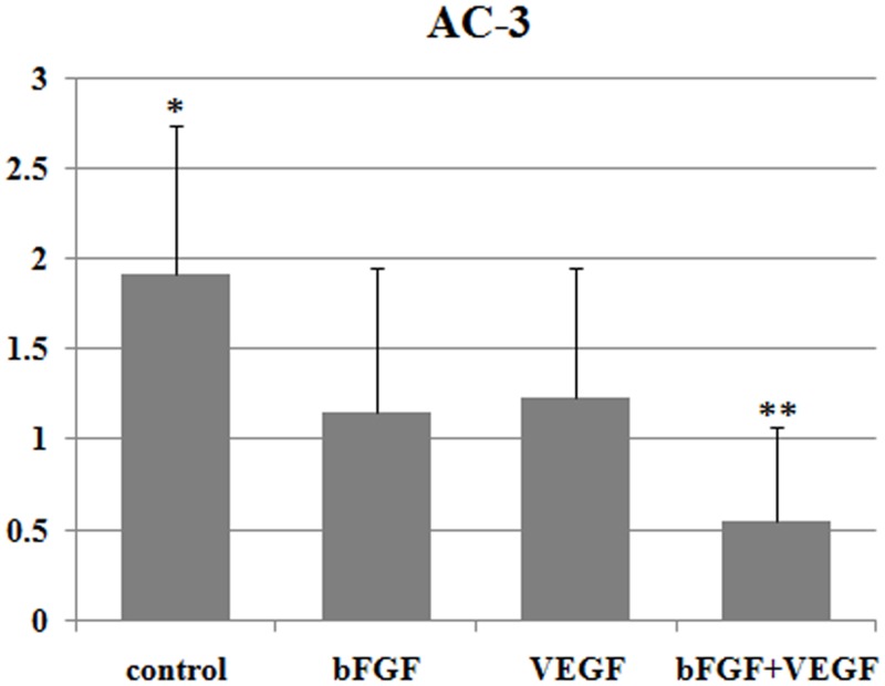
Fig 4. Apoptosis expression in grafts 7 days after grafting.
Results are presented as mean ± SD. Apoptosis (AC-3) levels were graded according to intensity and quantity: 0 = no apoptosis; 1 = few apoptotic cells with low staining intensity; 2 = apoptotic cells and medium staining intensity; and 3 = many apoptotic cells and high staining intensity.*Significantly higher than bFGF treatment and VEGF treatment (P <0.05). **Significantly lower than bFGF treatment and VEGF treatment (P<0.05).

