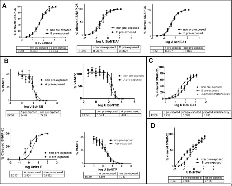
Fig 1. Sequential uptake of BoNTs.
Human iPSC derived neurons (iCell Neurons) (a, b, c) or primary mouse spinal cord cells (d) were exposed to the indicated BoNT serotypes (10 U / well BoNT/A, 1000 U / well BoNT/B, 1,000 U / well BoNT/D, 2,000 U / well BoNT/E, and 30 U / well BoNT/F) for 48 h to cause full SNARE cleavage (see S1 Fig). After complete removal of all extracellular toxin, the pre-exposed and not pre-exposed neurons were exposed to serial dilutions of the indicated BoNTs for 48 h in parallel, and cell lysates were analyzed for SNARE cleavage by Western blot using an anti-SNAP-25 antibody that recognizes both cleaved and uncleaved SNAP-25 equally and a VAMP2 antibody that detects only uncleaved VAMP2, which is quantitated in relation to syntaxin (all antibodies from Synaptic Systems). The graphs show the quantitative analyses of SNARE cleavage by the second BoNT serotype (as shown on the x-axis label), and the graph legends indicate the BoNT serotype added for pre-exposure. C: Cells first exposed to BoNT/E followed by exposure to BoNT/A were incubated for an additional 3 weeks before cell lysis to allow for recovery of BoNT/E cleaved SNAP-25. In all cases, each data point was measured in triplicate, and data were analyzed for statistical significance using an F-test in PRISM 6 software (n = 3).

