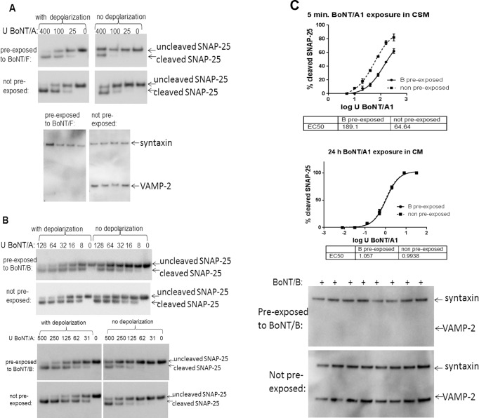
Fig 2. Activity dependent sequential BoNT entry into neurons.
Neuronal cultures were pre-exposed to BoNT/B or BoNT/F for 48 h, followed by complete removal of extracellular toxin. The pre-exposed or not pre-exposed cells were then exposed to the indicated concentrations (U / well) of BoNT/A1 in culture media or cell stimulation media (containing 56 mM KCl and 2.2. mM CaCl2) for 5 min, followed by removal of toxin, washing of the cells, and incubation for 24 h to allow for SNAP-25 cleavage. Cell lysates were analyzed for SNARE cleavage by Western blot and densitometry. All samples were tested in triplicate. (a) Human iPSC derived neurons (iCell Neurons) pre-exposed to 30 U BoNT/F. A Representative Western blot of SNAP-25 cleavage in depolarized (cell stimulation media) or not depolarized (culture media) cells is shown in the top figure, and VAMP-2 cleavage in BoNT/F pre-treated neurons in shown in the bottom figure. (b) Primary mouse spinal cord cells (top) or iCell Neurons (bottom) were exposed to 20,000 U BoNT/B / well (MSC cells) or 1000 U BoNT/B / well (iCell Neurons) for 48 h to cleave all VAMP2, followed by exposure to the indicated concentrations (U / well) of BoNT/A1 in culture media or cell stimulation media (containing 56 mM KCl and 2.2. mM CaCl2) for 5 min. Toxin was removed, cells washed and incubation for 24 hgig. to allow for SNAP-25 cleavage. Representative Western blots are shown. (c) Human iPSC derived neurons (iCell Neurons) pre-exposed to 1000 U BoNT/B for 48 h (bottom image), and after toxin removal cells were depolarized with 5 sequentially rounds of depolarization in cell stimulation media before exposure to serial dilutions of BoNT/A1 in either cells stimulation media for 5 min, or in culture media for 24 h. The graphs depict quantitative data from triplicate samples, respectively, and statistical significance was evaluated using an F-test in PRISM6.

