
Fig 4. Cytotoxicity of sequential exposure of human neurons to different BoNT serotypes.
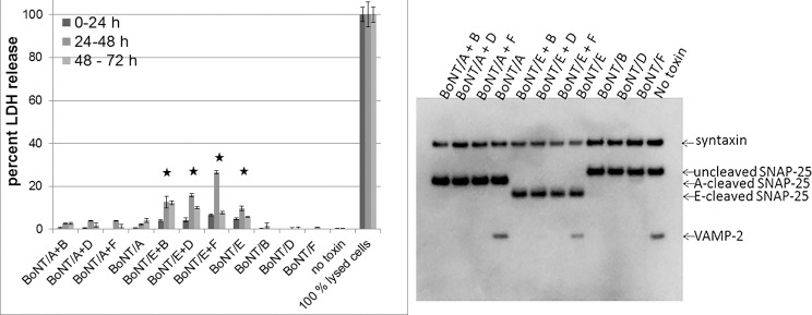
Human iPSC derived neurons (iCell Neurons) were exposed to 5 nM BoNT/A or BoNT/E for 24 h, followed by sequential exposure to 5 nM BoNT/B, /D, or /F for 24 h. Culture media was examined at 0–24 h, 24–48 h, and 48–72 h post addition of the second toxin for LDH release indicating cytotoxicity. Lysis buffer for the 100% lysed cells was added at 0 h (for the 0–24 h time point), 24 h (for the 24–48 h time point), and 48 h (for the 48–72 h time point). Media was replaced between each time point. All samples were measured in triplicate, and the average and standard deviation are shown in the graph. Statistical significance in relation to the control (cells with no toxin added) was determined by students t-test. Values that were significantly different at all three time points are indicated by a * (p < 0.01). One set of cells was lysed at 24 h post addition of the second toxin, and cell lysates were analyzed for SNARE cleavage by Western blot. One representative Western blot of triplicate samples is shown.