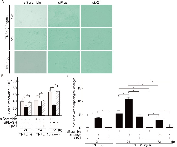
Fig 9. Effects of p21 and FLASH under TNF-α stimulation on cellular senescence in MCs.
(A) SA-β-gal staining at 48 h after knockdown of p21 and FLASH in MCs in the absence or presence of TNF-α shows induction of senescence. Representative data from one out of five independent experiments are shown. (B) The number of cells was determined microscopically using a hemocytometer at the indicated conditions. (C) Cells were counted on the basis of their morphological changes and expressed as the percentage of the total cell population. Results represent as arbitrary units and are shown as mean values ± SDs of at least three independent experiments. *P<0.001.

