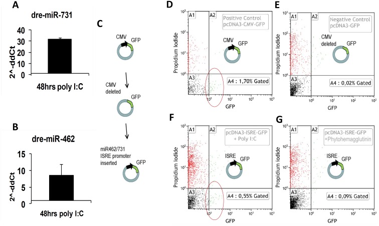
Fig 4. Expression of miR-462 and miR-731 can be activated by the TLR3 ligand poly I:C.
(A-B) Rainbow trout liver cells RTL-W1 in culture up-regulated miR-731 and miR-462 following stimulation with poly I:C. (C-G) Transfection with a mir-462 cluster promoter- GFP-reporter plasmid construct cells followed by stimulation with poly I:C, induced GFP expression in rainbow trout RTS-11 cells (F). Red circles in flow cytometry diagrams indicate cells with green fluorescence higher than the gated negative cells. Cells transfected with a positive control GFP-reporter plasmid containing the CMV promoter (D) and a negative control plasmid GFP reporter without any promoter (E) were used to gate for GFP positive cells (quadrant A4) and GFP negative cells (quadrant A3) respectively. Red dots (quadrant A1) and green dots (quadrant A2) indicate dead cells according to their staining above background with propidium iodide. Such cells were not considered in the analysis. No up regulation was seen in cells following stimulation with the TLR-4 ligand phytohaemagglutinin (PHA) (G).

