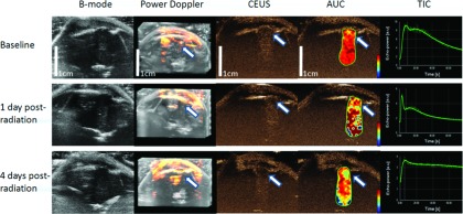
FIG. 3.

Changes in 2D B-mode, 3D power Doppler, CEUS, AUC, and TIC acquired at the different time points (baseline, 1 day postradiation, and 4 days postradiation) for a rat with a U87 glioma. The white arrows in power Doppler, CEUS, AUC are corresponding to the region showed homogeneous perfusion at baseline but with inhomogeneous and decreased perfusion at 1 day and 4 days postradiation. TIC also showed decreased perfusion for the contoured region of interest. Both 1 day postradiation and 4 days postradiation images share the same scale bar as in the baseline images.
