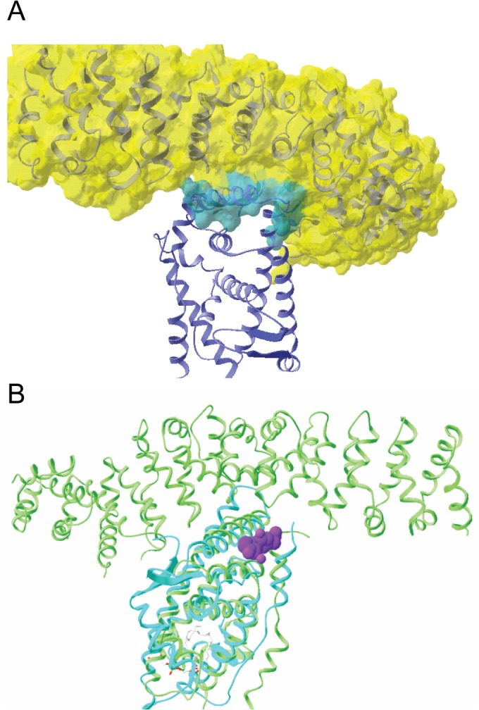
Fig 1. The Predicted β-Catenin Binding Site on AR is Near the Putative FKBP52 Regulatory Surface.

(A) Structure of the complex of nuclear receptor LRH-1 with β-catenin, PDB ID 3tx7. β-catenin is shown with a semi-transparent surface in yellow revealing its secondary structure elements as ribbons. LRH-1 is shown with blue ribbons for its secondary structure. The interfacial surface of these two molecules is shown in teal on the β-catenin surface. (B) Flufenamic acid from PDB ID 2PIT is shown in purple spheres as it binds to the androgen receptor shown with its secondary structure as teal colored ribbons. The Cα coordinates of androgen receptor in 2PIT were superimposed with the Cα coordinates of LRH-1 in 3TX7.
