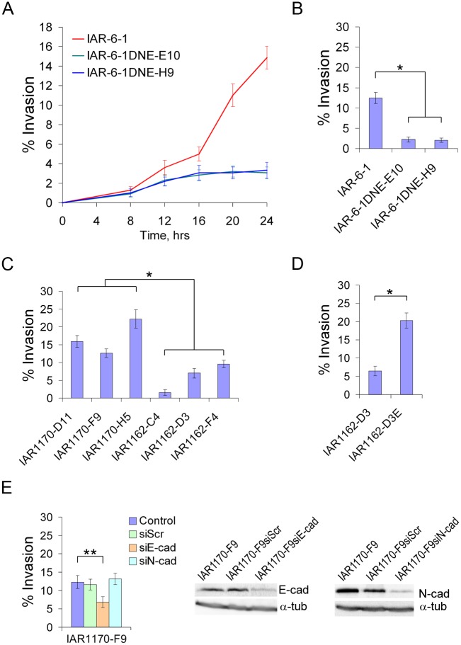
Fig 7. Invasive behavior of transformed cell lines.
(A) Dynamics of transepithelial migration of IAR-6-1 and IAR-6-1DNE cells. The diagram shows the percentage of transformed cells that invaded the IAR-2 monolayer and spread on the glass substrate below the monolayer to the number of seeded cells at various time points (mean ± SEM, n = 40). Transfection of a dominant-negative mutant of E-cadherin dramatically decreased the invasion of the epithelial monolayer by transformed cells. (B-D) A comparative study of the invasive behavior of a panel of transformed IAR cells in transepithelial migration assay. The diagrams show the percentage of transformed cells that invaded the IAR-2 monolayer and spread on the glass substrate to the number of seeded cells by 20 hours after seeding (mean ± SEM, n = 30). Asterisks indicate statistically significant differences (Kruskal-Wallis test, *—p‹0.001; **—p‹0.05). (B) IAR-6-1 and IAR-6-1DNE cells stably expressing a dominant-negative mutant of E-cadherin that abolished adhesive cadherin-based interactions. (C) Ras-transformed IAR1170 and IAR1162 clones. Cells that could form E-cadherin-based AJs (IAR1170-D11, IAR1170-F9, IAR1170-H5, IAR1162-D3E) were significantly more invasive than cells that could not (IAR1162-C4, IAR1162-D3, IAR1162-F4). (D) IAR1162-D3 cells and IAR1162-D3E cells stably expressing exogenous E-cadherin. (E) Effect of depletion of E-cadherin or N-cadherin by siRNA on transepithelial migration of IAR1170-F9 cells expressing both E- and N-cadherin.

