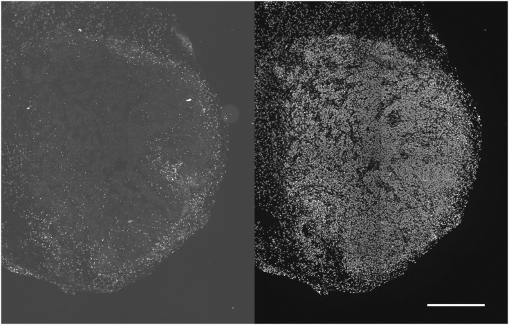
Fig 5. CD11b cells in SQ20B subcutaneous tumors.
CD11b positive cells were minimally found in the tumor interior (left panel). Total cells illustrated in the right panel, showing Hoechst 33342 nuclear staining (scale bar 0.5 mm). The lower density of nuclei along the left edge of the section showed the transition from tumor to normal tissue.

