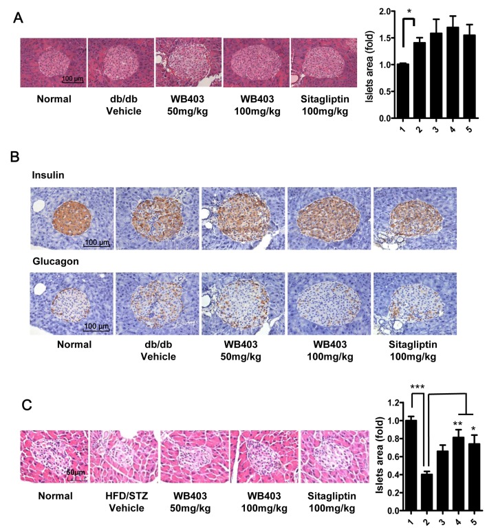
Fig 5. WB403 preserved the mass of pancreatic β-cells and normal distribution of α and β-cells.
(A) H&E staining of pancreas from db/db mice, and statistical result. Islets were sized by the Image J analysis software on alternated pancreatic sections spaced each by 100 μm. (B) Immunohistochemical analysis of pancreatic sections by anti-insulin antibody or anti-glucagon antibody. (C) H&E staining of pancreas from HFD/STZ mice, and statistical result. Results are representative islets from each group. 1–5 in the column graph on the right represents normal mice, diabetic mice treated with vehicle, WB403 50 mg/kg, WB403 100 mg/kg, sitagliptin 100 mg/kg respectively (n = 5). *p<0.05, **p<0.01, ***p<0.001 vs. diabetes-vehicle group.

