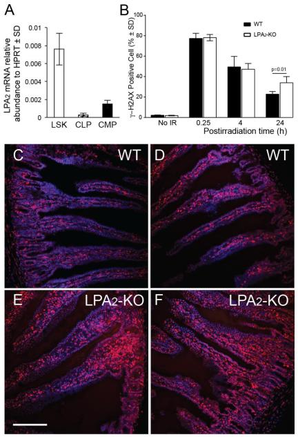
Figure 5. The role of LPA2 in DDR of HE stem/progenitor cells and in the jejunum.
Panel A. lpa2 expression detected by qRT-PCR analysis on stem (LSK) and lymphoid (CLP) and myeloid progenitor cell populations (CMP) from murine bone marrow. Panel B. LPA2-KO mice are deficient in the DDR process. Bone marrow isolated from mice exposed to 6 Gy TBI was subjected to flow cytometric analysis 15 minutes, 4 and 24 h postirradiation to determine γH2AX positive cells. LPA2-KO mice show elevated residual γH2AX compared to WT mice 24 h after 6 Gy total body irradiation. Representative of two experiments, n= 5 mice. *Denotes p value < 0.05 calculated by Student’s t-test. Panel C. γH2AX immunostaining in the jejuni of WT and LPA2-KO mice 4 h after 6 Gy total body irradiation shows elevated γH2AX expression in LPA2-KO mice compared to WT (γH2AX foci are stained red, nuclei are blue). Calibration bar = 250 μm.

