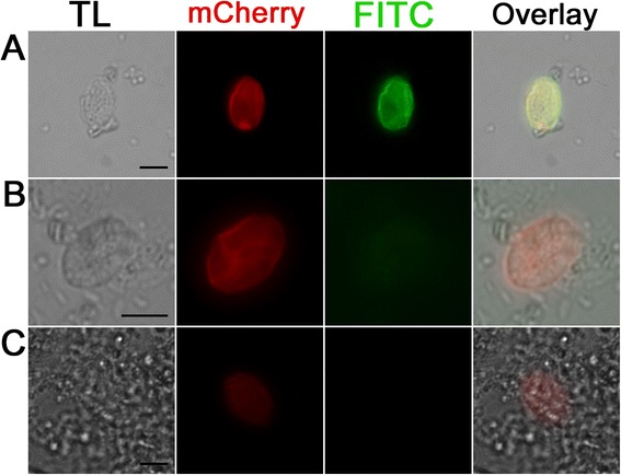
Fig. 6.

Immunofluorescence detection of mCherry in the sporocyst wall of mCherry positive but YFP negative sporocysts with goat anti-mCherry antibody and secondary anti-goat FITC antibody. a: Methanol fixed and Triton treated sporocyst incubated with primary anti-mCherry antibody and secondary anti-goat FITC antibody shows the signal of mCherry and the FITC signal of the antibody interaction with the sporocyst. b: Same antibody combination like in a but without Methanol fixation and Triton treatment shows similar mCherry signal, but a much reduced FITC signal. c: Methanol fixed sporocyst with Triton X100 treatment, without primary (anti mCherry) antibody but with secondary anti-goat FITC antibody does not show green fluorescent signal (K). Scale bar 5 μm. TL: Transmitted light micrograph, mCherry: mCherry signal of the sporocyst FITC: Signal of the anti-Goat FITC Antibody
