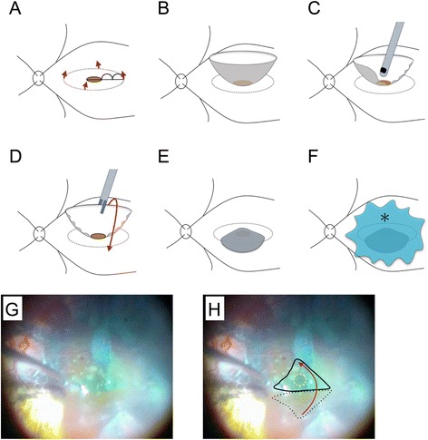
Fig. 2.

Diagrammatic representation of the modified inverted internal limiting membrane (ILM) flap technique and intraoperative photographs of the patient’s left eye. a After performing a 25-gauge micro-incision vitrectomy, the ILM was peeled off in a circular fashion. Red arrows show the direction of ILM peeling. The radius of the area of ILM peeled off was approximately 2.5 times the diameter of the macular hole (MH). b The ILM was not removed completely from the retina and remained attached to the edge of the MH. c We then trimmed half of the peeled ILM using a vitreous cutter, to facilitate the inversion of the ILM. d, e The remnant ILM was inverted with intraocular forceps to cover the entire MH. f The inverted ILM flap was covered with 1 % low molecular weight hyaluronic acid (asterisk) to stabilize it. Finally, we performed fluid-air exchange, but left the hyaluronic acid in the eye. g Intraoperative photograph showing the inverted ILM flap. h Explanatory drawing of (g), showing the area of the ILM flap before inversion (black dotted line), how we inverted the ILM flap (red arrow), the entire MH (white dotted line), and how we covered it with the inverted ILM flap (solid black line)
