Fig. 3.

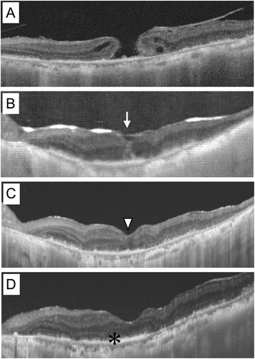
Swept source optical coherence tomography images showing the results of the inverted ILM flap technique. a The full-thickness large MH, which was 569 μm in diameter at the initial visit. b Three days after surgery, the image showed closure of the MH (arrow) in the gas-filled eye. c Twelve days after the surgery, the foveal contour had further improved (arrow head). d Although the MH had successfully closed 6 month after the surgery, leaving a U-shape depression, the ellipsoid zone remained defective (asterisk) and the visual acuity of the left eye was 20/400
