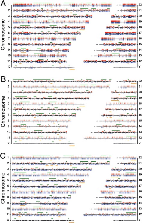
Fig. 2.

Chromosomal distributions of total mutations. a All LOHs and GOHs detected from AluScans of thirty cancer samples. b LOHs and GOHs detected from the whole genome sequences of a lung-to-liver metastatic cancer and its white blood cell control determined by Ju et al. [8]. c LOHs and GOHs detected from the whole genome sequences of a primary liver cancer and its normal liver tissue control determined by Ouyang et al. [9]. LOHs are shown as red vertical bars above cytobands, and GOHs as blue vertical bars below cytobands. The locations of common and rare fragile sites [19] are represented by horizontal green lines above, and horizontal orange lines below, the cytobands respectively. The chromosomal locations of all LOH and GOH sites are listed in Additional files 8: Table S5 and Additional files 5: Table S6
