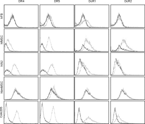
Figure 1.

Expression of DR4, DR5, DcR1 and DcR1 on the surface of foetal MSC, WJ cells (UMCS), adult bone marrow MSC, primary fibroblasts and Colo205 colon carcinoma cells. Cells in the log phase of their growth were harvested by trypsinization and labelled with DR4, DR5, DcR1 and DcR2-specific antibodies as described in Materials and Methods. The level of receptor expression was analysed by flow cytometry. The figure shows overlaid histograms of isotype control antibody (black line) and TRAIL-receptor antibody (grey line) labelled samples.
