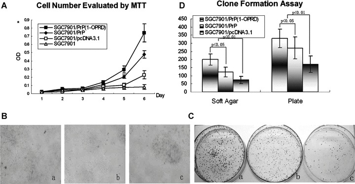
Figure 3.

Effects of PrPC (1-ORPD) on cell proliferation of gastric cancer cells. (A) Detection of the cell growth rate in vitro. Cell number was evaluated by the absorbance at 490 nm in MTT assay at the indicated time. The value shown is the mean of three determinations. (B) Detection of the clone formation in soft agar. Cell were placed in media containing soft agar and incubated for 20 days. The number of foci > 100 μm was counted. (a) SGC7901/PrPC (1-OPRD); (b) SGC7901/PrPC and (c) SGC7901/pcDNA3.1. (C) Detection of the clone formation in plate. Cell were placed in media containing plate and incubated for 20 days. The number of foci > 100 μm was counted. (a) SGC7901/PrPC (1-OPRD); (b) SGC7901/PrPC and (c) SGC7901/pcDNA3.1. (D) Detection of clone formation. Vertical bars represent mean ± S.D. from at least three separate experiments, each conducted in triplicate.
