
Figure 6.
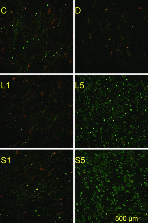
Apoptotic staining. Monolayers were stained with propidium iodide and fluorescein-labelled annexin V after 18 days of culture. Propidium (red) stains the nuclei of necrotic cells only. Green stain labels both apoptotic and necrotic cells. Therefore, apoptotic cells are single-stained (green), and necrotic cells are double-stained. Viable cells are not visible on these images. C, non-induced control; D, dexa; L1, 1 μM lovastatin; L5, 5 μM lovastatin; S1, 1 μM simvastatin; S5, 5 μM simvastatin.