
Figure 1.
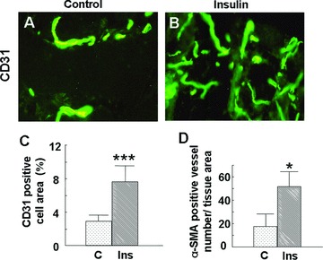
Subcutaneous injection of insulin stimulates angiogenesis in mouse skin: C57BL/6J mice (n= 6) were injected with insulin (0.03 U/20 μl saline) or saline (20 μl) subcutaneously at symmetric sites on the dorsum every 24 hrs for 5 days, and skin samples from the injected areas were collected at day 6. Tissue slides were stained with either anti-CD31 (PECAM-1) or anti-α-SMA, followed by FITC-conjugated goat antirat or goat antimouse secondary antibodies. Staining was visualised and pictures were taken using a Nikon immunofluorescence microscope (Bethesda, ML, USA). (A, B) Representative immunofluorescence images of CD31 staining: (A) control, (B) insulin; magnification 40×. Insulin injection results in the formation of longer blood vessels with more branches. (C) Percentage of CD31-positive cell area: the fraction of CD31-positive cell area relative to the total tissue area of each high-power field was measured using ImageJ software. At least eight fields per group were analysed. Insulin increases the percentage of the CD31-positive cell area. Data are shown as the mean value ± SD. Statistics indicate differences between the treatment and control. ***P < 0.001 versus control. (D) The number of blood vessels that express α-SMA were counted using fluorescence microscopy, and the vessel number was normalised by dividing by the total tissue section area. Insulin increased the number of α-SMA-positive blood vessels. Data are shown as the mean value ± SD. Statistics indicate differences between the treatment and control.*P < 0.05 versus control.