
Figure 2.
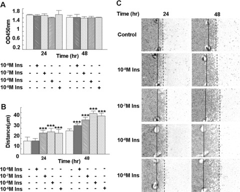
Insulin stimulates microvascular endothelial cell migration, but not proliferation, in a time- and dose-dependent manner. (A) HMEC-1 cells seeded in the 96-well tissue culture plates (1.0 × 10⁴ cells/well) were subjected to treatment with different concentrations of insulin for 24 or 48 hrs in the presence of BrdU. After fixation, the cells were processed according to the protocol of the manufacturer. Data are shown as the mean value ± SD. There were no statistically significant differences in endothelial cell proliferation following insulin treatment. (B, C) Cells were plated within cloning rings, the edges were marked and then the cells were treated with 10−8 M, 10−7 M, 10−6 M or 10−5 M insulin, respectively. Migration distance was measured at 24 and 48 hrs after treatment. Each treatment group was performed in triplicate. Data are shown as the mean value ± SD. Statistics are shown as comparisons between the treatment and control. **P < 0.01. Insulin significantly increased HMEC-1 migration, and the effect of insulin on cell migration correlated with the dose of insulin.