
Figure 3.
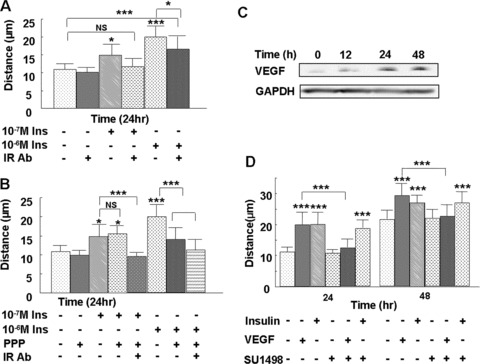
Insulin stimulates microvascular endothelial cell migration in an insulin receptor-dependent and a VEGF-independent manner. Endothelial cells were plated for the cloning ring migration assay, as described in Fig 2. (A) Cells were pre-treated with 1.5 μg of the neutralising insulin receptor Ab 29B4 for 1 hr, and then treated with 10−7 M or 10−6 M insulin for 24 hrs. The neutralising insulin receptor Ab completely inhibited 10−7 M insulin-induced cell migration and only partially inhibited 10−6 M insulin-induced cell migration, showing that the effect of 10−7 M insulin on endothelial cell migration is mediated primarily through the insulin receptor. (B) Cells were pre-treated with either 50 nM IGF-1 receptor inhibitor picropodophyllin or a combination of 1.5 μg of the insulin receptor Ab 29B4 and picropodophyllin for 1 hr, then treated with 10−7 M or 10−6 M insulin or left untreated for 24 hrs. The effects of the higher insulin concentration are mediated by both the insulin receptor and the IGF-1 receptor, whereas lower insulin concentrations are mainly mediated by the insulin receptor alone. (C) Cells were treated with 10−7 M insulin for the indicated time-points, the cell culture medium was then collected and VEGF levels were detected by Western blot. Insulin stimulates endothelial cell VEGF secretion. (D) Cells were either left untreated or pre-treated with 8 nM VEGFR inhibitor SU1498 for 1 hr, followed by treatment with 10−7 M insulin or 20 ng/ml VEGF; migration distance was measured at 24 and 48 hrs after treatment. Insulin-induced migration was not prevented with the VEGFR inhibitor, whereas VEGF-induced migration was prevented with this inhibitor. Each treatment group was performed in triplicate. Data are shown as the mean value ± SD. Statistics are shown as comparisons between the treatment and control, unless otherwise indicated. ***P < 0.001.