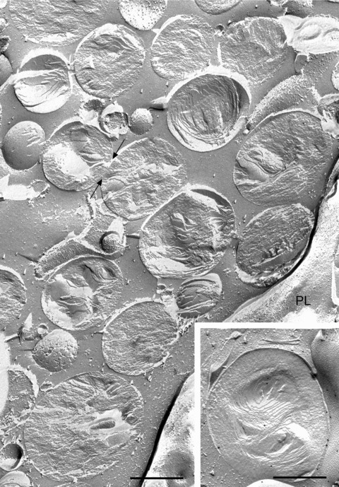
Figure 1.

Freeze-fracture view of typical appearance of cytoplasmic lipid droplets in THP-1 macrophages. The cells were incubated with 50 μg/ml acetylated LDL for 24 hrs to stimulate lipid droplet accumulation. The fracture plane frequently follows along the plane of organized lipids to give convex or concave views of the droplet; as the fracture path skips back and forth between lipid layers of the droplet, an ‘onion-like’ morphology is revealed. Other lipid droplets are cross fractured, revealing a stack of lipid layers in the core. In either case, the boundary of each droplet is usually clearly defined, demarcating one droplet from the next. Only occasionally do side-by-side droplets show regions of continuity (arrows), raising the possibility of an ongoing fusion event. PL, plasma membrane. In some instances, two stacks of lipid layers, each approximately ovoid in overall shape, are found within a single droplet, creating the impression that two droplets have somehow combined to make one (inset). In this example, the droplet has been immunogold labelled for adipophilin. Bar: 0.5 μm.
