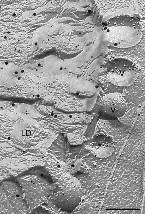
Figure 3.

Vesicular structures (v) at the periphery of a large lipid droplet. These might represent a mechanism for shedding excess phospholipid monolayer though how a stable bilayer vesicle could be created in such a situation is unclear. Alternatively, such structures may be involved in delivery to the droplet. In this example, the droplet has been double immunogold labelled for adipophilin (18 nm gold) and TIP 47 (12 nm gold). Bar: 0.2 μm.
