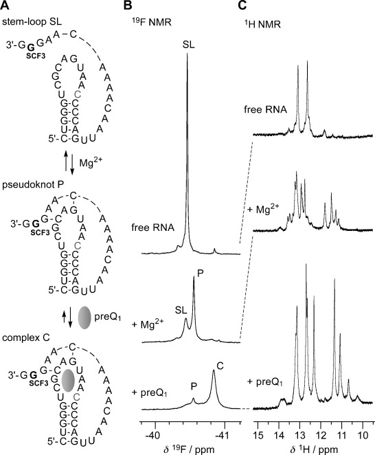
Figure 4.

NMR spectroscopic analysis of Mg2+-assisted RNA pseudoknot formation, and subsequent stabilization through binding of a small ligand (Thermoanaerobacter tengcongensis preQ1 class-I riboswitch), using a 2′-SCF3 guanosine label. A) RNA secondary structure model, B) corresponding 19F NMR spectra, and C) imino proton 1H NMR spectra. Conditions: cRNA=0.3 mM, 25 mM sodium cacodylate, pH 7.0, 298 K; additions: cMg2+=2.0 mM; followed by cpreQ1=1.2 mM. The cytosine that forms a Watson–Crick base pair with the preQ1 ligand is highlighted in grey.
