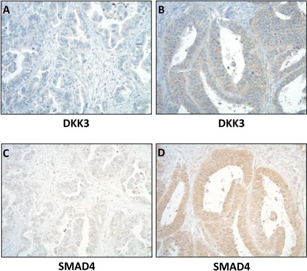
Figure 1.
Representative sections from a tissue microarray showing low (A) and high (B) cytoplasmic DKK3 staining in two esophageal adenocarcinomas. Moderate to high expression (2-3+) was found in 46.8% (29/62) of esophageal adenocarcinomas. Low (C) and high (D) SMAD4 staining correlated with DKK3 in the same tumors. Original magnifications are all 100x.

